
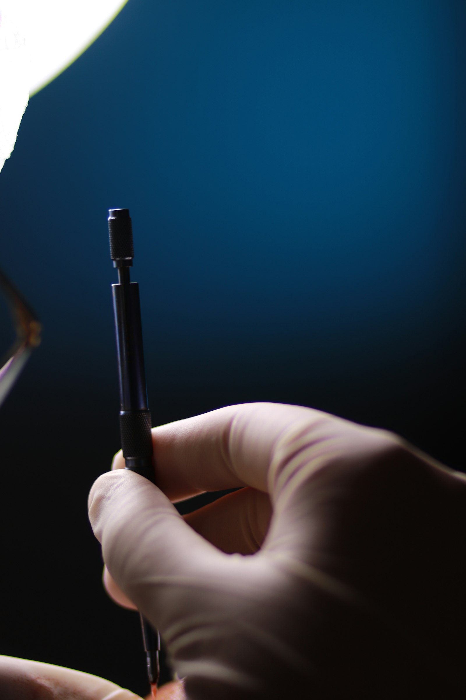
Case Discussion: Microsurgical Nerve Repair After Sharp Laceration
15 May 2026 · Surgeons & Fellows
Presentation
A sharp laceration to the volar surface of a finger, with reported numbness distal to the injury and no active bleeding requiring urgent control. Clinical exam is used to localise the likely level of nerve injury and assess for concurrent flexor tendon involvement, which frequently coexists with digital nerve lacerations at this location.
Decision points
Timing of repair. Clean, sharp lacerations with minimal contamination are generally suitable for primary repair at the time of initial surgery. Contaminated wounds, crush components, or significant nerve gap may warrant staged management.
Repair technique. For a clean laceration with minimal gap, direct epineural or fascicular repair under magnification is standard. Where tension-free approximation isn't achievable, options include nerve conduits for short gaps or nerve grafting for longer defects — the threshold for each varies by surgeon and gap length, and is worth discussing explicitly with trainees rather than treated as automatic.
Magnification and instrumentation. Operating microscope versus loupe magnification is case-dependent; fascicular-level repair generally benefits from microscope use, particularly in mixed motor-sensory nerves.
Post-operative protocol
Protective splinting to avoid tension on the repair, followed by a structured, therapist-guided desensitisation and sensory re-education programme once early healing allows. Recovery of protective sensation typically precedes fine discriminative sensation, and patients should be counselled on this realistic timeline from the outset.
Teaching point
The technical repair is often the more straightforward part of these cases. The judgement calls — repair versus graft, timing, and setting expectations for sensory recovery — are where case discussion adds the most value for trainees.